


Upholding a patient-centric philosophy, our professional cardiology team strives to serve the community amid the imbalance between public and private healthcare. Early treatment is critical for recovery. Understanding that many patients hesitate to seek private cardiac surgery due to cost concerns and end up waiting long in the public system, Hong Kong Asian Heart Centre offers reasonable, fully transparent pricing to deliver affordable and reliable cardiac care.
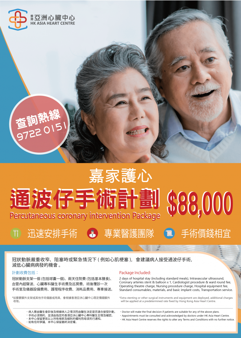
Beyond cardiac check-up packages, we provide comprehensive cardiac surgery plans, including coronary intervention, minimally invasive mitral/tricuspid valve surgery, and renal sympathetic denervation. No public hospital referral is required, and the plans are open to all citizens. All-inclusive fees cover specialist surgical charges, ward rounds, basic operating room and equipment costs, hospitalization expenses, and post-surgery follow-up consultations, eliminating patients’ concerns about unexpected extra charges.