
Many people mistakenly believe all heart diseases cause the same severe symptoms. In fact, heart disease encompasses various conditions including coronary heart disease and arrhythmia, which present different signs — some even have no early warnings.
Heart disease is among Hong Kong's top three causes of death. Its incidence is rising and affecting more young people. Everyone should understand the risks and take preventive measures early.
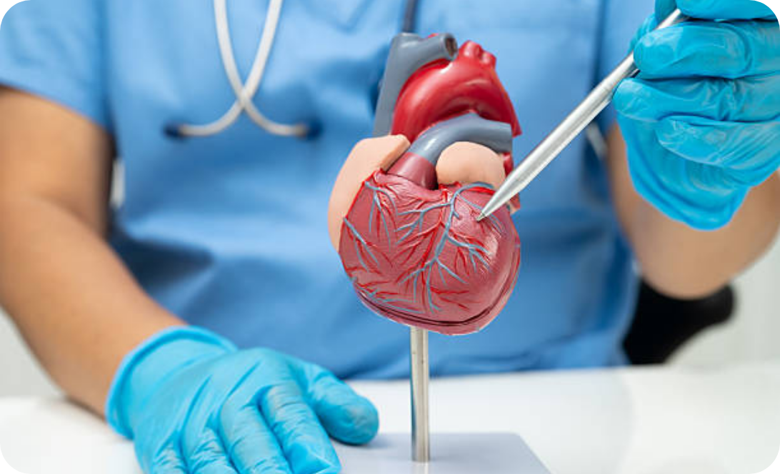
According to government statistics, around 10.9 people die from coronary heart disease on average each day in 2022, making it the leading cause of death among all heart diseases. Coronary heart disease develops when the coronary arteries become narrowed, blocked or hardened. This impedes blood flow and reduces blood supply to the heart, resulting in cardiac hypoxia and triggering heart problems. During an episode, patients often experience symptoms such as angina pectoris. An acute myocardial infarction may cause irreversible damage to heart muscle, and can lead to sudden cardiac death in severe cases. Some patients have no warning signs before their first attack, leaving them unprepared.
Arterial narrowing, blockage and hardening are the direct causes of coronary heart disease, mainly linked to fat accumulation and ageing. Nevertheless, the condition is increasingly affecting younger people in recent years. Below are common contributing factors:

Angina pectoris is one of the most common symptoms during early attacks. Actual manifestations vary depending on the degree of arterial blockage and the patient’s other chronic illnesses. The following are typical early symptoms of coronary heart disease:
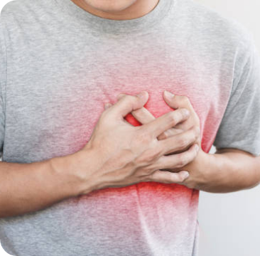
It is usually triggered by physical exertion or emotional excitement. Patients experience chest pain and tightness. In severe cases, discomfort or pain may spread to the jaw, neck, upper abdomen, arms or back. Symptoms generally ease within 15 minutes of rest or after taking medication.

Insufficient blood supply to the brain during an attack may cause dizziness or even fainting in severe cases
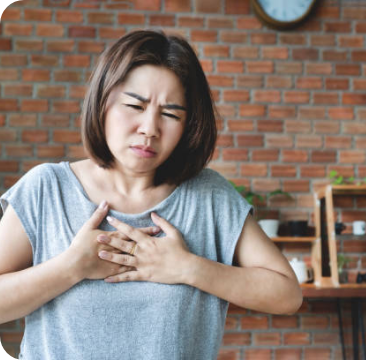
Patients may suffer from breathing difficulties due to inadequate cardiac blood output

Coronary heart disease may impair cardiac function, causing persistent tiredness
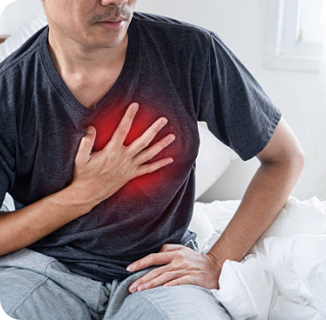
Some patients develop abnormal heart rhythms, which may deteriorate into ventricular fibrillation and cause death within minutes

Diabetic patients, the elderly and women may have subtle warning signs. Medical consultation is recommended once suspicious symptoms appear. Since some people have no symptoms at all before an attack, regular health check-ups are essential.
At rest, a normal adult’s heart rate ranges from 60 to 100 beats per minute. Arrhythmia refers to abnormally fast, slow or irregular heartbeats. It is mostly associated with structural heart problems and can be classified into several types:
1. Ventricular Fibrillation
Ventricular fibrillation is the leading cause of sudden cardiac death and the most dangerous type of arrhythmia. During an episode, ventricular muscles contract rapidly and erratically, disrupting normal heartbeat. The heart fails to pump blood effectively, leading to cerebral hypoxia. Patients may lose consciousness and die within minutes.
2. Atrial Fibrillation
Atrial fibrillation is the most common type. Disordered electrical signals make the atria escape the control of the sinoatrial node, resulting in rapid and irregular contractions and impaired cardiac pumping function. Blood may stagnate inside the atria and form clots. If clots enter the bloodstream, they may block blood vessels and cause stroke. Many patients have no obvious symptoms, which increases hidden risks.
3. Ventricular Tachycardia
This condition arises from ventricular abnormalities. Patients experience a rapid heartbeat, sometimes exceeding 200 beats per minute. Episodes are often brief, lasting less than 30 seconds with no obvious warning signs, so they are easily overlooked.
4. Sick Sinus Syndrome
Dysfunction of the sinoatrial node causes irregular and bradycardic heart rhythm, or even brief cardiac arrest. It is commonly seen in the elderly due to ageing, but may also occur in younger people affected by other heart diseases.
5. Supraventricular Tachycardia
Abnormal electrical signals at the atrioventricular junction trigger sudden rapid heartbeat that subsides abruptly. Attacks may last from minutes to hours. Most cases are congenital with no early symptoms that only manifest in adulthood. Some cases develop secondary to other heart conditions.
Ventricular fibrillation is the leading cause of sudden cardiac death and the most dangerous type of arrhythmia. During an episode, ventricular muscles contract rapidly and erratically, disrupting normal heartbeat. The heart fails to pump blood effectively, leading to cerebral hypoxia. Patients may lose consciousness and die within minutes.
Atrial fibrillation is the most common type. Disordered electrical signals make the atria escape the control of the sinoatrial node, resulting in rapid and irregular contractions and impaired cardiac pumping function. Blood may stagnate inside the atria and form clots. If clots enter the bloodstream, they may block blood vessels and cause stroke. Many patients have no obvious symptoms, which increases hidden risks.
This condition arises from ventricular abnormalities. Patients experience a rapid heartbeat, sometimes exceeding 200 beats per minute. Episodes are often brief, lasting less than 30 seconds with no obvious warning signs, so they are easily overlooked.
Dysfunction of the sinoatrial node causes irregular and bradycardic heart rhythm, or even brief cardiac arrest. It is commonly seen in the elderly due to ageing, but may also occur in younger people affected by other heart diseases.
Abnormal electrical signals at the atrioventricular junction trigger sudden rapid heartbeat that subsides abruptly. Attacks may last from minutes to hours. Most cases are congenital with no early symptoms that only manifest in adulthood. Some cases develop secondary to other heart conditions.
There are numerous high-risk factors for arrhythmia, many of which are related to other cardiovascular diseases and personal lifestyle:
Below are three common warning signs of arrhythmia:
Tachycardia (Rapid heartbeat). Patients may experience shortness of breath, palpitations, chest pain, chest tightness or dizziness. Severe cases such as ventricular fibrillation may lead to loss of consciousness and sudden cardiac death.
Bradycardia (Slow heartbeat). Patients feel weak, fatigued, dizzy and short of breath. Severe cases may cause confusion or even sudden death.
Irregular heartbeat. The heart beats erratically with skipped beats while the overall rate stays within normal range. Patients may feel chest tightness, palpitations or dizziness. Severe cases can result in shock or sudden death.
Some patients have mild or unnoticeable symptoms, while others remain completely asymptomatic and are only diagnosed incidentally during routine check-ups.

The human heart has four valves that control blood flow and prevent backflow. The mitral valve and tricuspid valve regulate blood flow between the atria and ventricles on the left and right sides respectively. Valvular heart disease refers to lesions and impaired function of heart valves, leading to stenosis, prolapse, sclerosis or even rupture.
A diseased valve such as one with calcification cannot open fully, narrowing the blood passage and limiting blood supply. The heart has to work harder to pump blood, which may eventually lead to heart failure.
Valves normally close tightly to prevent blood regurgitation. Incomplete closure causes blood to flow backward, known as valvular regurgitation. This condition may result in cardiomegaly, heart failure or infection.
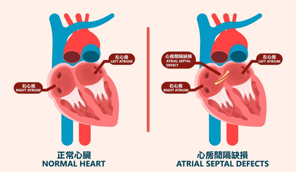
As the name suggests, congenital heart diseases are present at birth and mainly caused by structural heart defects. Many patients have no obvious symptoms in childhood and are only diagnosed during adult health check-ups or after an acute episode. Atrial septal defect and ventricular septal defect are two common types, collectively known as heart shunt.
An atrial septal defect is a hole in the wall separating the left and right atria, while a ventricular septal defect refers to a gap between the left and right ventricles.
Since these conditions are congenital, major risk factors include non-modifiable elements such as genetic inheritance.
Small holes may close naturally without causing any symptoms. Unclosed defects allow excessive blood to flow into the pulmonary artery and raise pressure inside pulmonary vessels. Early warning signs may include shortness of breath, lower limb oedema, abdominal distension and easy fatigue. Advanced cases can lead to pulmonary congestion and heart failure. Long-term pressure damage to pulmonary vessels may also cause pulmonary hypertension.
The primary function of heart muscle is to pump blood. Cardiomyopathy refers to heart disease caused by abnormalities in the heart muscle. Due to genetics or other factors, the myocardium may become thinned, thickened or stiffened. These changes impair normal cardiac contraction and blood pumping, weakening the heart. Severe attacks can lead to heart failure.
The ventricles become enlarged while the myocardium remains normal or thinned. The weakened heart muscle cannot pump sufficient blood. This condition may result from coronary artery disease, valvular heart disease, drug or alcohol abuse, viral infections or hereditary factors.
The heart muscle thickens due to ageing, high blood pressure or genetics. Thickened myocardium reduces the size of heart chambers and lowers cardiac output.
This is a rare type more commonly seen in elderly patients. It is linked to cardiac scar tissue, amyloidosis, hemochromatosis, sarcoidosis or complications after radiotherapy. The myocardium turns stiff and loses elasticity, preventing the heart from proper expansion and filling, which in turn affects heartbeat.
Another rare form mainly caused by genetics, congenital right ventricular abnormalities or myocarditis. The right ventricular tissue is replaced by scar tissue or fat, causing abnormal electrical conduction. An acute episode may trigger severe arrhythmia and even sudden cardiac death.
Early warning signs may be subtle, and some patients experience no symptoms before a sudden attack. Below are common manifestations:
Heart failure occurs when the heart weakens and loses its ability to pump and supply blood effectively. Early warning signs may be vague. As the condition worsens, pulmonary oedema is likely to develop, which may further lead to liver and kidney failure and greatly increase health risks.
Symptoms of heart failure are often subtle. Some patients only realize they are ill when an attack happens, missing the optimal time for treatment.
Long-term high blood pressure can damage blood vessels and the heart:
Persistent high blood pressure constricts blood vessels. If the coronary arteries are affected, it may result in coronary heart disease.
High blood pressure forces the heart to pump harder, which gradually thickens the heart muscle over time.
Chronic hypertension may deform heart valves and impair their normal opening and closing function.
Infections caused by bacteria, viruses, fungi or parasites may lead to heart conditions such as myocarditis and endocarditis.
Warning signs: fever, chills, joint or muscle pain, fatigue, nausea, difficulty breathing, shortness of breath and unexplained weight loss

It is divided into resting and ambulatory types. This non-invasive test records the heart's electrical activity.

A non-invasive examination that uses ultrasound to capture real-time clear images of the heart, helping specialists assess cardiac structure and function.

Also known as coronary angiography, it mainly evaluates calcification and stenosis of the coronary arteries.

It delivers high-quality images and enables comprehensive analysis of cardiac structure and function in one scan.

An invasive procedure used to assess blood vessel conditions such as stenosis and blockage.

It checks indicators such as cholesterol and triglycerides to evaluate the risk of heart disease.
Medication: For patients with mild conditions, medicines such as anticoagulants can reduce the risk of heart attacks. Doctors may also prescribe drugs to relieve symptoms including oedema and chest pain.
Surgical Treatment: Surgery can improve or cure heart diseases according to specific conditions. Common procedures include coronary angioplasty, coronary artery bypass grafting, catheter ablation, heart valve replacement and pacemaker implantation.
As the name implies, congenital heart diseases are present at birth and mainly result from structural abnormalities of the heart. Some patients have no obvious warning signs or mild symptoms during childhood, and the condition is only detected during adult health check-ups or after an acute episode. Atrial septal defect and ventricular septal defect are two common types.
An atrial septal defect refers to a hole in the wall separating the left and right atria, while a ventricular septal defect is a gap between the left and right ventricles. Collectively, these conditions are known as heart shunt.
Since these disorders are congenital, major risk factors are non-modifiable, such as genetic inheritance.
Small holes may close naturally without causing any symptoms. If the defect remains unclosed, excessive blood will flow into the pulmonary artery and raise pressure in pulmonary blood vessels. Early signs may include shortness of breath, swelling of the lower limbs or abdomen, and easy fatigue. Advanced heart shunt may lead to pulmonary congestion and heart failure. Long-term pressure damage to pulmonary vessels can also cause pulmonary hypertension.
Heart disease has consistently ranked among the top three causes of death in Hong Kong's annual mortality registrations, with coronary heart disease being the leading fatal category, predominantly affecting males. According to government statistics, in 2022, an average of approximately 10.9 people died from coronary heart disease every day, accounting for nearly 60% of relevant deaths. Moreover, coronary heart disease is showing a trend of onset at a younger age.
As the causes of coronary heart disease are closely linked to modern lifestyle habits, regular physical check-ups and coronary heart disease screenings are crucial for prevention. Additionally, if patients receive early intervention upon the onset of early coronary heart disease symptoms, more safe and effective treatment options will be available.
Every heartbeat propels blood circulation, delivering nutrients and oxygen to all parts of the body through the bloodstream. The heart muscle, which works around the clock, also requires a blood supply to function properly. The arteries responsible for supplying blood to the heart are located on its surface; their distribution resembles a crown, hence they are known as coronary arteries. Coronary heart disease is mainly caused by the narrowing, blockage, or hardening of the coronary arteries, which restricts or blocks blood flow. This reduces the blood supply to the heart, causing myocardial hypoxia, which can damage or even kill the heart muscle, resulting in coronary heart disease.
Narrowed, blocked, or hardened blood vessels directly lead to coronary heart disease. Apart from some non-modifiable factors, the root cause of the disease is the accumulation of fat and aging of blood vessels.
The accumulation of fat in the arteries narrows the vessel lumen and impairs blood flow. In severe cases, "fatty plaques" form, further narrowing or completely blocking the arteries—a condition known as atherosclerosis. When this occurs in the coronary arteries, it causes coronary heart disease.
Since fat accumulation in blood vessels is directly related to obesity, obesity is widely recognized as a major cause of coronary heart disease, and weight loss is a common requirement for patients during treatment. However, not all coronary heart disease patients are obese. Even slim individuals may develop the disease due to high blood lipids or metabolic disorders that cause fat buildup in blood vessels. Therefore, regardless of body type, anyone experiencing symptoms should seek medical attention promptly and undergo regular screenings.
The human body ages with advancing age. As vascular tissues age, they thicken and stiffen, narrowing the vessels and impairing cardiac blood flow—this is why coronary heart disease is more common in the elderly. However, age is not the only cause of vascular aging or coronary heart disease; unhealthy lifestyles, stress, and genetics can also harden blood vessels. With changes in modern living conditions and patterns, vascular aging is no longer exclusive to the elderly, and coronary heart disease is becoming increasingly prevalent among younger people.
In fact, coronary heart disease is mostly a chronic condition developed over years, and most causes are related to daily habits. Understanding these common risk factors is the first step to prevention.
High blood pressure constricts blood vessels, increasing long-term strain on arteries and the heart, accelerating atherosclerosis and leading to coronary heart disease.
Persistent high blood sugar damages blood vessel health, accelerates atherosclerosis, impairs cardiac blood supply, and causes coronary heart disease. Furthermore, high blood sugar may damage cardiac nerves, so some diabetic patients may not feel the most common symptom—angina pectoris—until the condition is severe. Regular screenings are therefore critical for diabetic patients.
Excess cholesterol in the blood accumulates in blood vessels, gradually causing narrowing and atherosclerosis, blocking cardiac blood flow and leading to coronary heart disease.
Obesity involves excess fat storage in the body; when fat accumulates in blood vessels, it causes coronary heart disease. Obesity also significantly increases the risk of diabetes, high blood pressure, and high cholesterol—all causes of the disease—making weight loss a common requirement in treatment.
Nicotine in cigarettes constricts blood vessels and raises blood cholesterol levels, causing atherosclerosis. High-risk individuals should quit smoking early to reduce their risk.
Many alcoholic beverages are high in starch and sugar; long-term excessive drinking easily triggers diabetes and high cholesterol, both risk factors for coronary heart disease.
Mental tension or excessive stress raises blood pressure, increasing cardiac strain over time and leading to coronary heart disease. Chronic severe stress can also cause anxiety disorders with symptoms similar to those of coronary heart disease—patients with concerns should seek medical help promptly to avoid delayed treatment.
Regular moderate exercise improves myocardial blood circulation and prevents vascular aging. It also helps maintain a healthy weight, lower blood pressure, control blood sugar, and relieve stress—all linked to coronary heart disease causes. For diagnosed patients, exercise addresses root causes and reduces the frequency of attacks.
Excess salt intake is directly linked to high blood pressure; too much sugar increases insulin production, leading to diabetes long-term; high-fat diets (especially saturated fats) accelerate fat accumulation and vessel blockage—all risk factors for coronary heart disease. A light, nutritious diet is vital in treatment.
Coronary heart disease is not directly hereditary, but a family history increases the risk significantly—relatives should pay extra attention to cardiovascular health. Those with a family history of other risk factors (e.g., diabetes) should also undergo regular screenings.
Female hormones help maintain vascular elasticity and regulate blood cholesterol levels, making men more susceptible to coronary heart disease. However, post-menopausal women experience a decline in female hormones, and aging is a risk factor—so older women’s risk rises to match men’s.
The body ages with time, and blood vessels are prone to aging, which can lead to coronary heart disease.
Angina pectoris is the most common early symptom of coronary heart disease, but actual symptoms depend on the extent of vessel blockage and the patient’s other chronic conditions. Acute myocardial infarction can cause heart muscle necrosis, with symptoms including severe chest pain, dizziness, difficulty breathing, arrhythmia, and a sharp drop in blood pressure—even sudden death in severe cases. Below are common symptoms:
1. Angina Pectoris:
Coronary heart disease results from blocked blood flow to the heart via coronary arteries, causing myocardial ischemia and angina—a classic symptom. It is usually triggered by heavy meals, physical activity, or emotional excitement. Patients feel chest pain, tightness, heaviness, compression, or a burning sensation. In severe cases, discomfort radiates to the jaw, neck, upper abdomen, arms, or back. Symptoms typically subside within 15 minutes of rest or medication.
2. Dizziness:
During an attack, insufficient blood supply to the brain causes dizziness; severe cases lead to fainting.
3. Shortness of Breath / Difficulty Breathing:
Impaired cardiac blood supply causes this symptom.
4. Fatigue / Limb Weakness:
Declining heart function leads to persistent tiredness.
5. Palpitations:
Some patients experience arrhythmias; severe cases lead to ventricular fibrillation, which can be fatal within minutes.
6. Cold Sweats, Nausea and Vomiting:
Symptoms are often subtle in diabetic patients, the elderly, and women—some have no obvious warning signs and are only diagnosed via screening. Anyone with suspected symptoms should consult a doctor immediately to avoid delayed treatment.
1. Blood Test:
Fat accumulation and vessel narrowing are core causes. Blood tests measure cholesterol, triglycerides, and other markers to assess disease risk. They are common, affordable, and included in standard check-ups. Patients with abnormal results may need further screening.
2. Resting Electrocardiogram (ECG):
A non-invasive, quick, and simple test included in most check-up packages. Patients lie flat while the device records the heart’s electrical activity at rest, helping detect arrhythmias, myocardial ischemia, or necrosis caused by coronary heart disease.
However, it only records short-term activity—no attack during the test means diagnosis may be missed, delaying treatment. It is not ideal for patients already experiencing symptoms.
3. Treadmill Exercise ECG:
Another non-invasive test. Patients walk on a treadmill with attached devices, with intensity gradually increased by medical staff. Blood pressure is monitored, and the test stops immediately if symptoms or abnormalities appear.
This screening diagnoses coronary heart disease and latent conditions, evaluates treatment effectiveness, and assesses physical fitness—especially cardiac function under stress. It is unsuitable for people with mobility difficulties.
4. Echocardiogram:
Non-invasive, providing clear real-time heart images to examine myocardial contraction, blood flow speed/direction, and assess cardiac structure and function. Ischemia from coronary heart disease affects myocardial activity, guiding further testing if needed.
Safe and convenient for regular check-ups, but it cannot visualize coronary arteries—unsuitable for obese patients, those with emphysema, or suspected cases.
5. Computed Tomography (CT) / Coronary Angiography:
Atherosclerosis and blockage are key causes. Non-invasive CT coronary angiography effectively checks coronary artery lumen narrowing and calcification, providing accurate data for risk assessment, diagnosis, and early treatment. It also evaluates outcomes after stenting or bypass surgery.
6. Cardiac Catheterization and Coronary Angiography:
This test combines cardiac catheterization (assessing heart structure/function) and coronary angiography (evaluating vessel narrowing/blockage) using X-rays. Under local anesthesia, a catheter is inserted into blood vessels and guided to the heart. Contrast dye is injected incrementally to capture X-ray images, enabling precise assessment of disease severity and optimal treatment for complex cardiac structures.
It is invasive with some risks but highly accurate and critical for acute cases or before angioplasty, as it can help open blocked arteries.
1. Medication Treatment:
Medications target the root issues: blocked/hardened vessels and myocardial ischemia. Nitroglycerin (sublingual tablets) relaxes and dilates coronary arteries to boost blood supply, rapidly relieving angina. Beta-blockers and calcium channel blockers lower blood pressure and reduce cardiac strain to prevent angina. Antiplatelet drugs (including aspirin) prevent blood clots that block coronary arteries.
Since high blood pressure, high cholesterol, and diabetes are causes, doctors prescribe relevant medications to control the patient’s condition.
2. Angioplasty (Stenting):
A common invasive treatment performed under local anesthesia. Using cardiac catheterization, a specialized balloon and stent are delivered to the blocked artery. The balloon expands to open the vessel, and the stent is implanted to maintain blood flow.
It is widely used for acute coronary syndrome, is minimally invasive (no open-chest surgery), has low trauma and a high success rate, making it an effective option.
3. Coronary Artery Bypass Grafting (CABG):
Used for severe vessel narrowing when angioplasty is ineffective. This major surgery requires general anesthesia and open-chest access. Healthy blood vessels are harvested from the patient’s body and grafted to bypass blocked arteries, restoring blood flow.
While it resolves severe blockages, it is highly invasive with greater risks—highlighting the importance of early screening and treatment to avoid advanced disease.
Arrhythmia (also known as Cardiac Arrhythmia)
Your heart rate naturally speeds up during exercise and remains steady at rest. However, if your heartbeat suddenly becomes too fast, too slow, or skips a beat without a reasonable cause, it may be a symptom of arrhythmia. Although arrhythmia is usually not immediately life-threatening, it doubles the risk of death, triples the risk of heart failure, and increases the chance of stroke by up to five times.
Fortunately, with advances in medical technology, there are more treatment options for arrhythmia than ever before. Therefore, if you have experienced suspected symptoms of arrhythmia, such as sudden rapid or slow heartbeat, you can first learn more about the causes, symptoms, and treatment plans of arrhythmia.
Arrhythmia refers to any abnormal heartbeat or heart rhythm disorder. What constitutes an abnormal heartbeat? First, at normal rest, the heart rate of a healthy adult is 60 to 100 beats per minute, maintaining a steady rhythm. Arrhythmia includes a heart rate below 60 beats per minute (bradycardia), above 100 beats per minute (tachycardia). In addition, even if the heart rate is within the normal range, an irregular, chaotic heartbeat is also classified as arrhythmia.
The heart has a complex structure, and its beating sustains blood supply to the entire body. Different structural problems lead to different types of arrhythmia, which directly determine the treatment plan selected by specialists. Below is a detailed classification:
1. Ventricular Fibrillation (VF) (Tachycardia):
This is the most dangerous and severe type of arrhythmia, as most sudden cardiac deaths are caused by ventricular fibrillation. During an attack, the ventricular muscles contract rapidly and irregularly, causing a chaotic, quivering heartbeat. As a result, the heart cannot pump blood effectively. When blood fails to reach body organs, the brain is deprived of oxygen, leading to loss of consciousness within seconds and eventual cardiac arrest. Without prompt treatment, the patient may die within minutes.
The causes of this arrhythmia include myocardial ischemia, heart failure, and coronary artery disease. Among them, myocardial ischemia is the most common cause—insufficient oxygen supply to the heart muscle triggers ventricular fibrillation.
2. Atrial Fibrillation (AF) (Irregular Heartbeat):
Atrial fibrillation is the most common type of arrhythmia. While not as immediately fatal as ventricular fibrillation, it can lead to serious conditions such as stroke and heart failure. Some patients experience obvious symptoms like irregular, extremely rapid heartbeat, while others have no symptoms at all, increasing hidden risks.
The upper chambers of the heart are the left and right atria. The "sinoatrial node" on the wall of the right atrium acts as a natural pacemaker, regulating the heartbeat. This arrhythmia occurs when abnormal heart cells emit chaotic electrical signals, causing the atria to quiver and contract rapidly and irregularly, out of the node’s control. This impairs the heart’s pumping function; blood may pool in the atria and form clots. If a clot enters the bloodstream, it can block vessels and cause diseases such as stroke.
3. Ventricular Tachycardia (VT) (Tachycardia):
This arrhythmia stems from abnormalities in the ventricles. Patients experience a rapid heartbeat, sometimes exceeding 200 beats per minute. (Episodes may be brief, lasting less than 30 seconds, with no other symptoms, so it is often overlooked.)
However, the danger of this arrhythmia should not be underestimated, as ventricular tachycardia can progress to more severe, fatal arrhythmias such as ventricular fibrillation (VF). Therefore, any heart rhythm abnormality requires timely testing and treatment as needed, even without severe symptoms.
4. Sick Sinus Syndrome (SSS) (Bradycardia):
Also known as sinus node dysfunction, this condition arises from abnormalities in the sinoatrial node—the natural pacemaker that generates and regulates the heartbeat. During an attack, patients experience irregular heartbeat, bradycardia, or even temporary cardiac arrest.
The main cause is aging, so it is more common in elderly patients. However, certain heart diseases can also affect the node’s function, meaning young people may also develop bradycardia due to sick sinus syndrome.
5. Supraventricular Tachycardia (SVT) (Tachycardia):
This is a relatively low-risk type of arrhythmia. During an attack, abnormal electrical signals disrupt the atrioventricular junction, causing sudden rapid heartbeat that stops abruptly. Episodes can last from minutes to hours.
Most cases are congenital, though symptoms may not appear until adulthood. Some cases develop later in life alongside other diseases. Symptoms include shortness of breath, palpitations, chest pain, and dizziness. While low-risk, untreated SVT can rarely lead to shock or sudden death.
Below are common symptoms of arrhythmia:
1. Tachycardia:
A resting heart rate exceeding 100 beats per minute. Patients may experience:
Shortness of breath or difficulty breathing
Palpitations
Chest pain or tightness
Dizziness
In severe cases, loss of consciousness or sudden death
Types causing tachycardia include ventricular fibrillation, atrial fibrillation, ventricular tachycardia, and supraventricular tachycardia.
2. Bradycardia:
A resting heart rate below 60 beats per minute. Patients typically feel:
Weakness
Persistent fatigue
Dizziness
Shortness of breath
In severe cases, confusion or sudden death
The main type causing bradycardia is sick sinus syndrome.
3. Irregular Heartbeat:
The heart rate is within the normal range but fluctuates erratically or skips beats. Patients may feel:
Chest tightness
Palpitations
Dizziness
In severe cases, shock or sudden death
The main type causing irregular heartbeat is atrial fibrillation.
Since the heart beats continuously all day, subtle symptoms are hard to detect. Some patients have no symptoms or warning signs and are only diagnosed during routine check-ups. Additionally, symptoms like shortness of breath, palpitations, or dizziness resemble those of anxiety or panic attacks, which may delay diagnosis and treatment. Therefore, anyone with suspected arrhythmia symptoms should consult a professional promptly to identify the cause and receive appropriate treatment.
Besides congenital factors in some patients, there are many high-risk factors for arrhythmia, many closely related to other cardiovascular diseases and personal lifestyle:
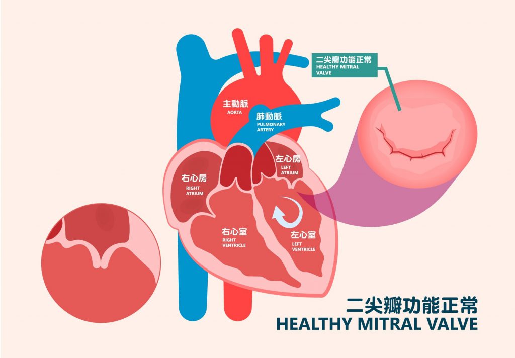
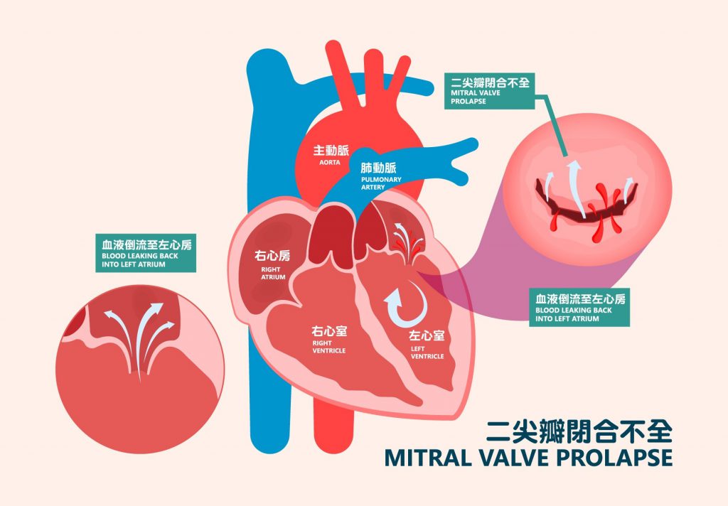
The mitral/tricuspid valves control the blood flow between the atria and ventricles. Each heart valve is composed of thin but strong tissue.
As blood flows between the atria and ventricles, the valves open and close naturally to allow blood to flow in the proper direction. However, if the valves fail to close completely, blood will flow backward in the opposite direction, which is known as valvular regurgitation. Valvular regurgitation can lead to heart failure and may cause arrhythmia, stroke, or even sudden cardiac death.
Atrial fibrillation (AFib) is a common type of arrhythmia. Patients usually experience an irregular and extremely rapid heartbeat.
AFib is classified into three main types:
● Paroxysmal (lasts no more than 7 days, typically within 24 hours)
● Persistent (lasts more than 7 days)
● Permanent (lasts over one year)
It is estimated that around one percent of the population in Hong Kong suffers from atrial fibrillation. Based on the current population of over 7 million, this accounts for approximately more than 70,000 patients.
The heart consists of the left and right atria in the upper chamber and the left and right ventricles in the lower chamber.
The sinoatrial node located on the upper wall of the right atrium acts as the heart's natural pacemaker. When abnormal cells in the atria generate erratic electrical impulses, the atria lose control by the sinoatrial node and contract rapidly and irregularly, resulting in fibrillation.
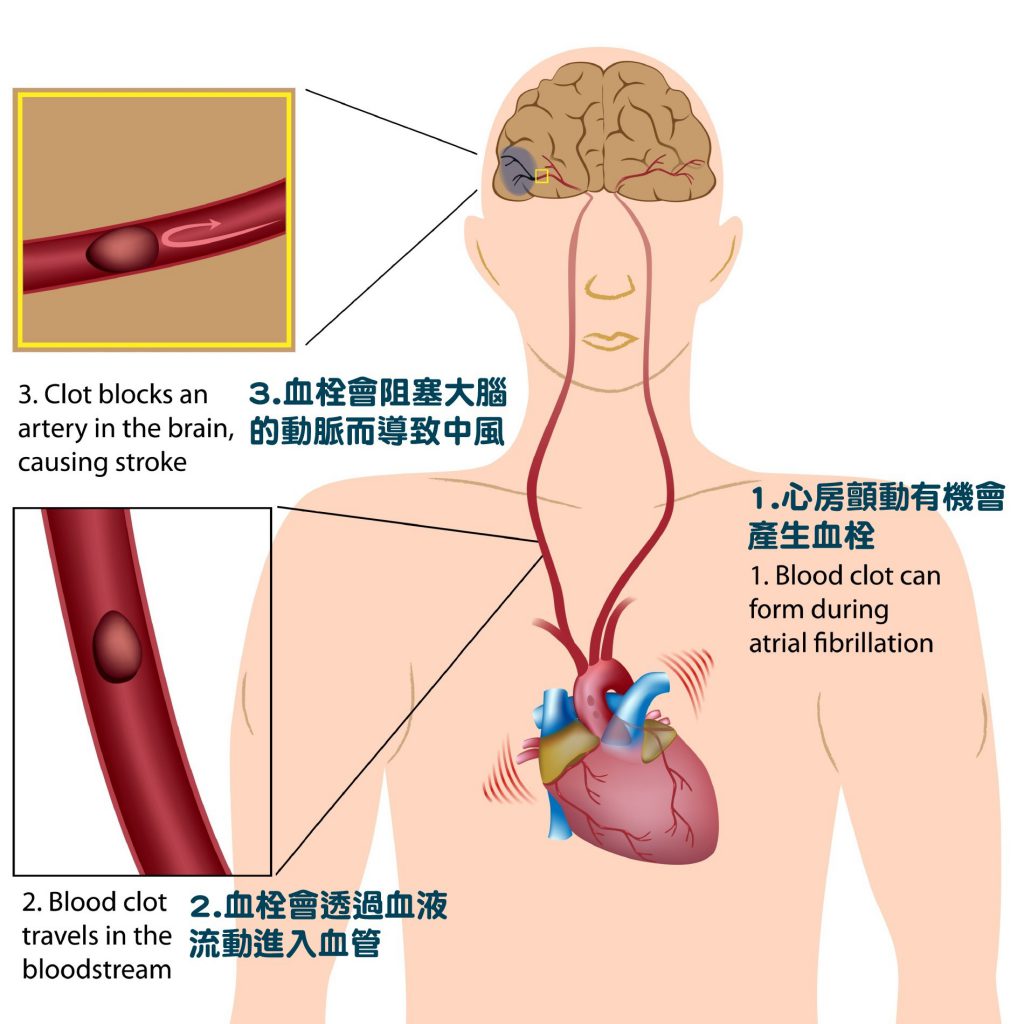
The ventricles fail to pump blood effectively to the whole body. Some blood may stagnate in the atria and form blood clots. If these clots enter the bloodstream, they can block blood vessels and lead to illnesses such as stroke, a common complication.
Atrial septal defect is a common congenital heart disease. A gap forms between the septum of the left and right atria, causing abnormal blood shunting and increasing the burden on the heart.
In addition, the foramen ovale is located between the left and right atria. It naturally closes in most people after birth; if it remains unclosed, it is called patent foramen ovale. Adults with this condition can live a normal life, but patent foramen ovale increases the risk of blood clot formation.