關於我們
中心簡介
我們的團隊
中心地址 / 交通資訊
暖心行動
暖心行動
查詢 / 報名
常見問題
相片集
醫療服務
推廣優惠
服務及收費
健康資訊
健康資訊
傳媒報導
個案分享
心臟病種類
病人須知
惡劣天氣下的診症安排
求診指南
常見問題
關於我們
中心簡介
我們的團隊
中心地址 / 交通資訊
暖心行動
暖心行動
查詢 / 報名
常見問題
相片集
醫療服務
推廣優惠
服務及收費
健康資訊
健康資訊
傳媒報導
個案分享
心臟病種類
病人須知
惡劣天氣下的診症安排
求診指南
常見問題
系統訊息
×
{{message}}
訂閱消息
×
電郵地址
歡迎光臨香港亞洲心臟中心
×
請選擇語言
中文
English
同心護您心
擁有多年治療心臟病的醫療團隊,秉持以心服務市民的精神,通過先進醫療技術及設備,為患者提供優質及全面的心臟專科服務。
閱讀更多
服務介紹
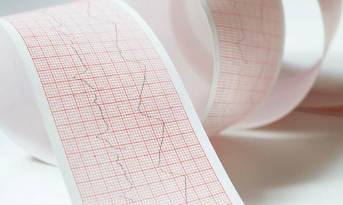
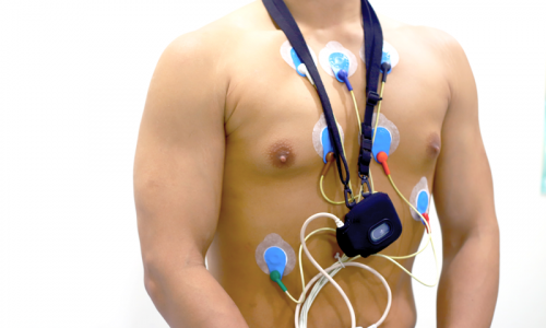
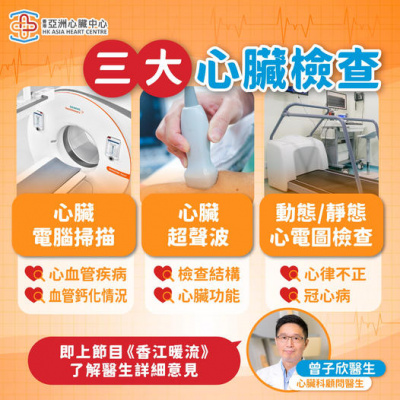
靜態心電圖
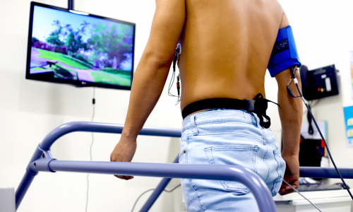
運動踏板心電圖
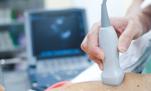
心臟超聲波
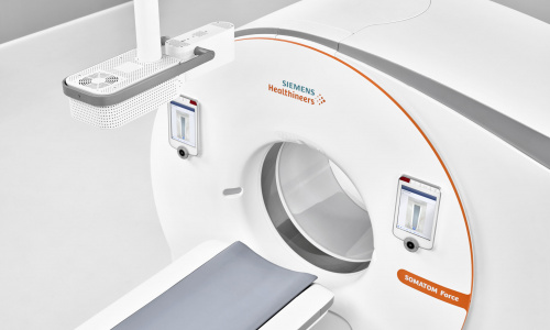
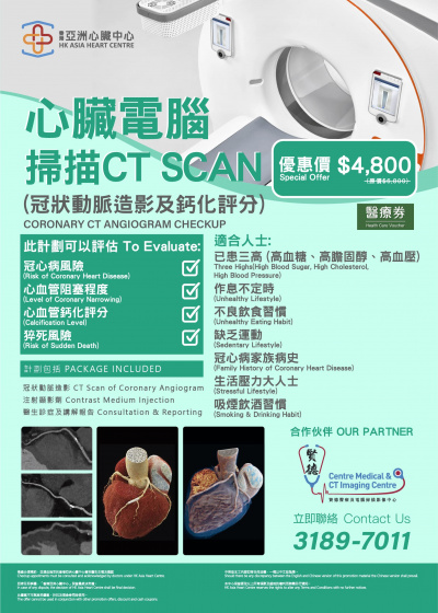
心臟電腦掃描
攜帶式活動心電圖
Kardia 移動心電圖儀
閱讀更多
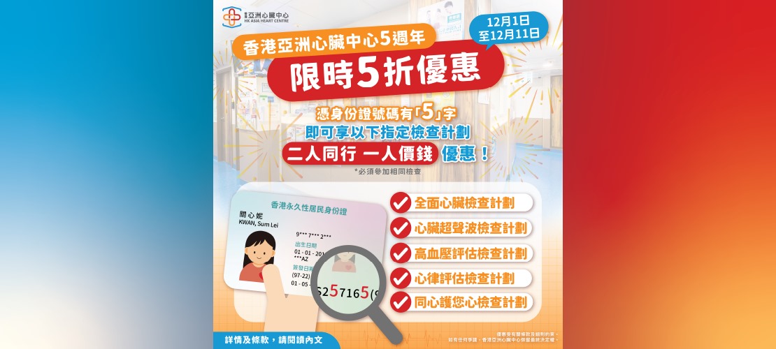
推廣優惠
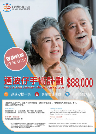
$88,000 通波仔手術計劃
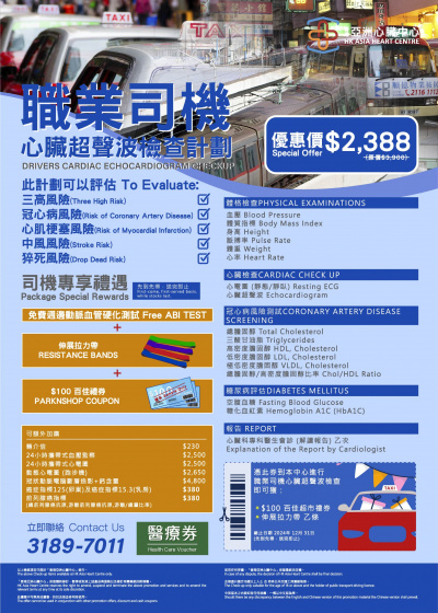
【職業司機心臟檢查計劃】
心臟電腦掃描 (CT SCAN)
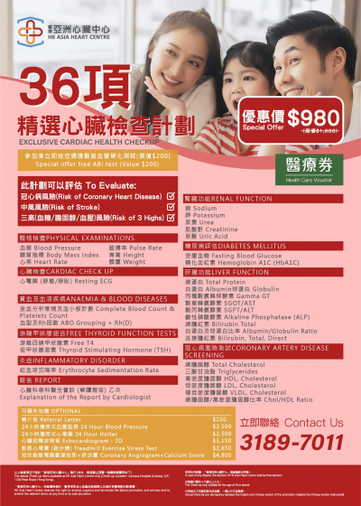
精選心臟檢查計劃
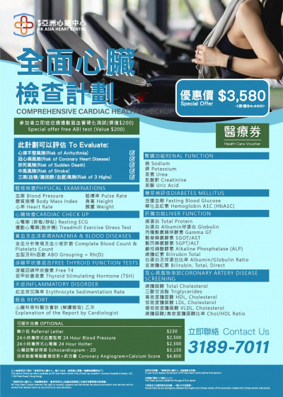
全面心臟檢查計劃
心臟超聲波檢查計劃
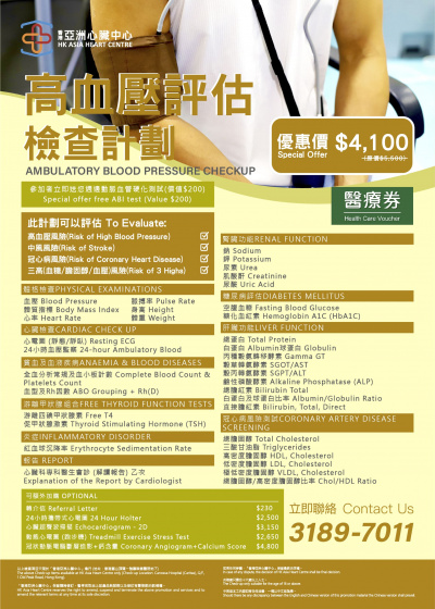
高血壓評估檢查計劃
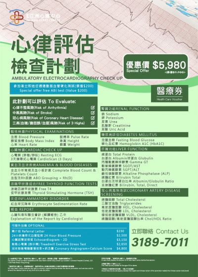
心律評估檢查計劃
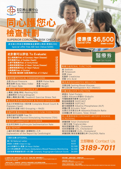
同心護您心檢查計劃
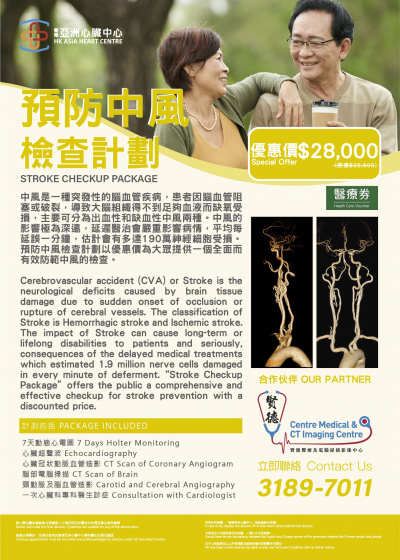
預防中風檢查計劃
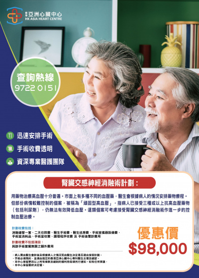
腎臟交感神經消融術計劃
心臟復康計劃
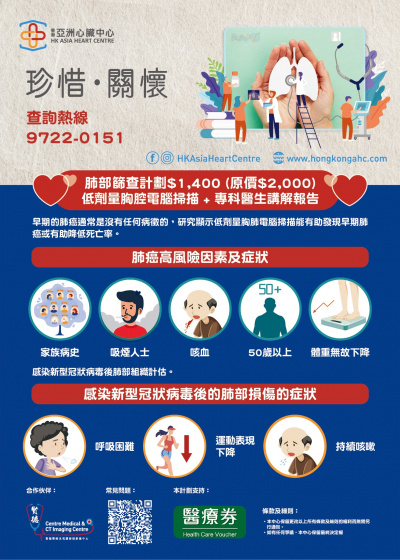
肺部篩查計劃
閱讀更多
健康資訊
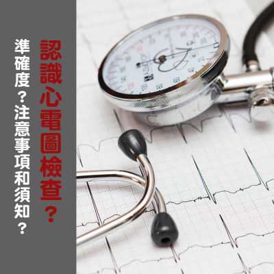
【心電圖的準確率高嗎? 有風險或副作用有嗎?】
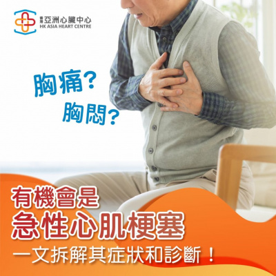
胸痛?有機會是急性心肌梗塞!
【健康一Heart | 復活蛋的秘密】
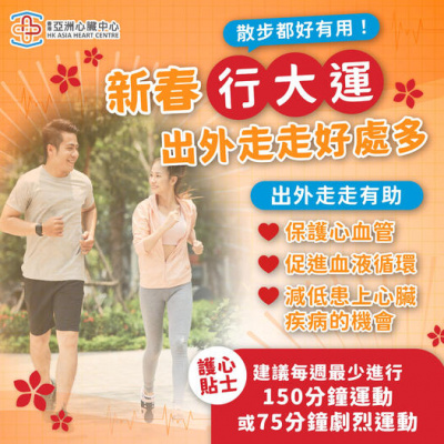
【新春行大運,出外走走好處多】
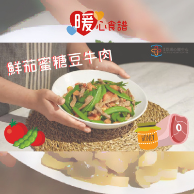
【暖心食譜|鮮茄蜜糖豆牛肉】
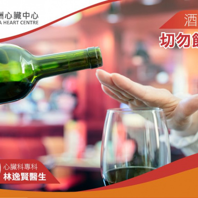
飲酒的風險
要揀啱自己適合嘅檢查計劃
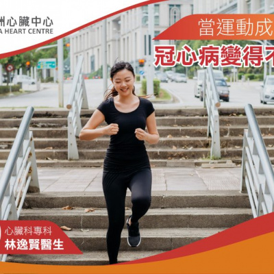
恆常的帶氧運動 助肥胖人士對抗冠心
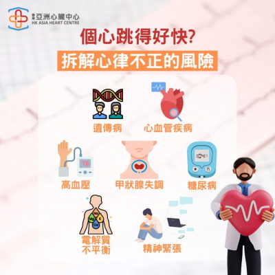
個心跳得好快?一文拆解心律不正的風險
【暖心食譜 | 香蕉藍莓蛋糕】
閱讀更多
傳媒報導
【中暑容易中獎難 器官衰竭仲麻煩】
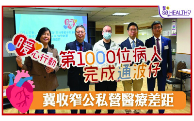
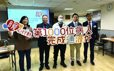
【公私營醫療協作未見進展 「暖心行動」助千人通波仔冀拉近差距】
疫境救命「暖心價」助患者免輪候「通波仔」
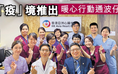
「疫」境推出暖心行動通波仔
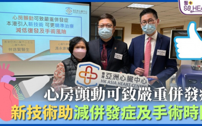
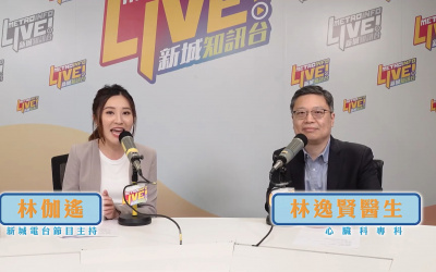
心房顫動可致嚴重併發症 新技術助減併發症及手術時間
【心房顫動增中風風險,香港亞洲心臟中心引入最新脈衝場消融術 (PFA)】
「暖心行動」助千人「通波仔」 最年輕31歲
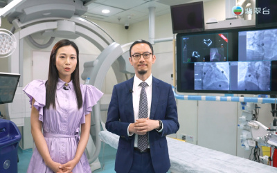
【醫+直播】心臟疾病手術迷思:通波仔和二次阻塞
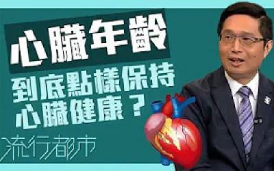
【流行都市:到底你個心臟喺幾歲?】
【健康護心師:心瓣疾病必定要做開胸手術?】
閱讀更多
個案分享
暖心重生‧沒有症狀的高血壓
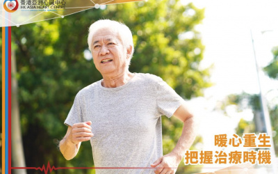
暖心重生 ‧ 把握治療時機
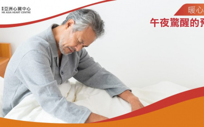
暖心重生‧午夜驚醒的預兆
暖心重生 ‧ 省卻半年的等待
暖心重生 ‧ 4次病發的經歷
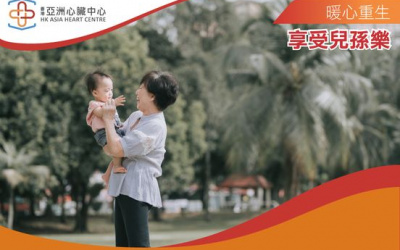
暖心重生.享受兒孫樂
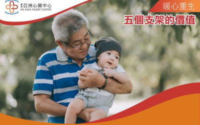
暖心重生.五個支架的價值
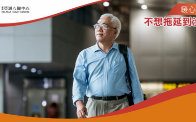
暖心重生‧不想拖延到沒救
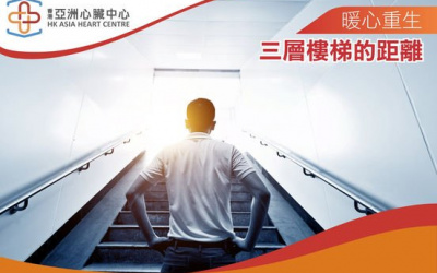
暖心重生・三層樓梯的距離
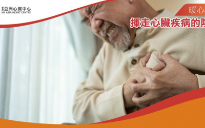
暖心重生‧揮走心臟疾病的陰影
閱讀更多